MicroCT
Contrast enhanced MicroCT
Recent work in our lab has investigated the use of contrast agents such as phosphotungstic acid (PTA) to investigate if it can bind preferentially to microstructural components of the vessel, such as collagen. In this study, porcine carotid arteries and human atherosclerotic plaques were stained with a concentration of 1%PTA staining solution and imaged using MicroCT to establish the in-situ architecture of the tissue and measure collagen content. A histological assessment of the collagen content was also performed from picrosirius red (PSR) staining.
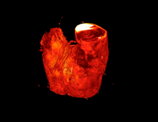
Figure 1 shows how staining the porcine carotid artery with PTA can show the individual layers of the vessel wall and from the overall HU, can further be segmented into its individual layer as required.
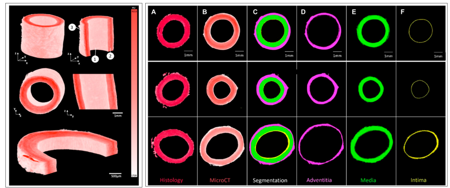
Figure (1): 3D render of porcine carotid artery stained for 15hours using 1% PTA solution. Each image shows different cross-sectional views highlighting the capability to visualize multiple layers of the vessel wall: (1) Intimal layer (2) Medial Layer (3) Adventitial layer. The second image here shows the segmentation of three PTA-stained porcine carotid arteries into their respective layers (A) Histological PSR image (B) PTA stained MicroCT image (C) Segmentation (D) Adventitia (E) Media (F) Intima.
Figure 2A shows a similar visualization of the MicroCT images to the 2x histology images. Furthermore, it was observed that regions with higher collagen content had higher Hounsfield Units, see Figure 2C. However, across samples there was no correlation between the Hounsfield Units and the collagen content within the vessel. It was observed that this was likely due to varying tissue volumes across the samples and the resulting variability in the intensity of the stain across different samples. Thus, the Hounsfield units were normalised with respect to volume. Once volume was accounted for, the collagen content observed across a number of tissue samples showed a positive moderate correlation with an r = 0.596 between the histology and MicroCT as shown in Figure 2D.
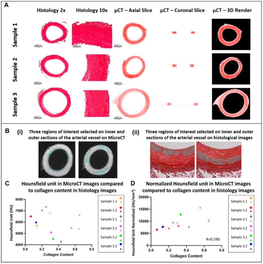
Figure (2): Qualitative and quantitative comparison of MicroCT to histology (A) Qualitative comparison observing the overall structure of porcine carotid arteries. (B) Region of interest selection for quantitative comparison of the collagen content observed in porcine carotid arteries. (C) Increase in collagen content observed in different sections of vessel wall whereby the external media present higher collagen content and HU then the internal media, internal (circles) and external (stars) regions of interest in the media. (D) Pearson correlation between HU from MicroCT and collagen content from PS-Red images when normalised with respect to volume.
Figure 3 shows that the staining protocol was successfully adapted to human atherosclerotic plaques. By leaving the sample in the PTA solution for a longer period of time, the samples could be imaged using the MicroCT and rendered in 3D. Furthermore, qualitative assessment revealed distinctive regions within the plaque, including the lumen, lipid rich cores, calcified regions and fibrous caps, as shown in Figure 3B.
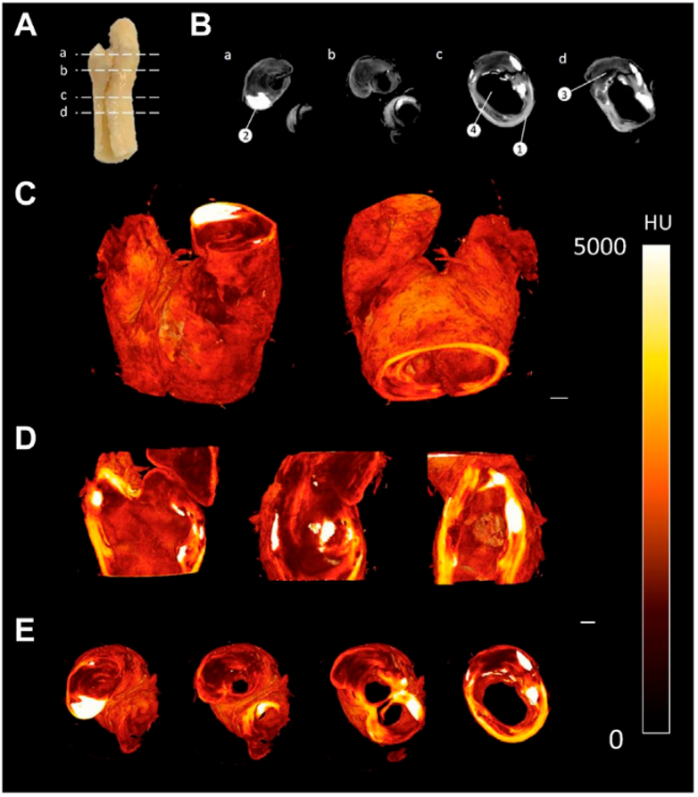
Figure (3): CEµCT of human atherosclerotic plaque (A) Slice locations in atherosclerotic plaque. (B) Axial cross-sections directly from MicroCT show multiple components within the tissue: (1) Outer wall of plaque, (2) Calcification, (3) Lipid, and (4) Lumen of plaque. (C) 3D render of atherosclerotic plaque. (D) Sagittal and coronal cross-sections through atherosclerotic plaque. (E) Axial cross-sections through atherosclerotic plaque. Scale bar = 1 mm.
Publications
A. Hanly, R.D. Johnston, C. Lemass, A. Jose, Phosphotungstic acid ( PTA ) preferentially binds to collagen- rich regions of porcine carotid arteries and human atherosclerotic plaques observed using contrast enhanced micro-computed, Front. Physiol. (2023) 1–10. https://doi.org/10.3389/fphys.2023.1057394.