ULTRASOUND
Studies in our lab also focus on performing mechanical testing to gain insights on the mechanical properties of arterial tissue. This subsequently could allow us to yield invaluable information on arterial diseases such as carotid atherosclerosis. More particularly, we combine the use of non-invasive ultrasound imaging to move towards clinically relevant features. Performing inflation testing allows to pressurize samples and reproduce physiological loading while keeping the tissue in its intact form.
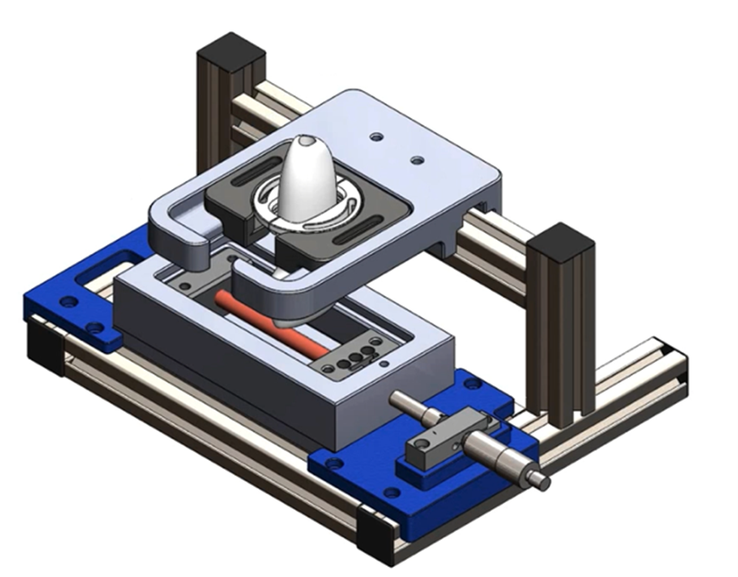
Figure 1: Inflation testing set-up combining non-invasive ultrasound imaging
Bi-layered diseased arterial tissue surrogates with different stenosis percentages and lipid-rich inclusion were developed using polyvinyl alcohol (PVA).

Figure 2: Photographs of phantoms (top row) and associated B-mode scans (bottom row)
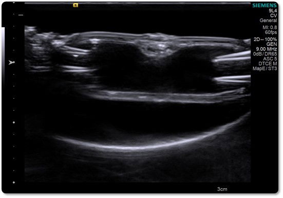
Figure 3: Longitudinal view of phantom with 25% stenosis
Inflation testing of those developed phantoms was successfully performed and allow us to gain a deeper understanding of diseased arterial tissue while mimicking in vivo loading.
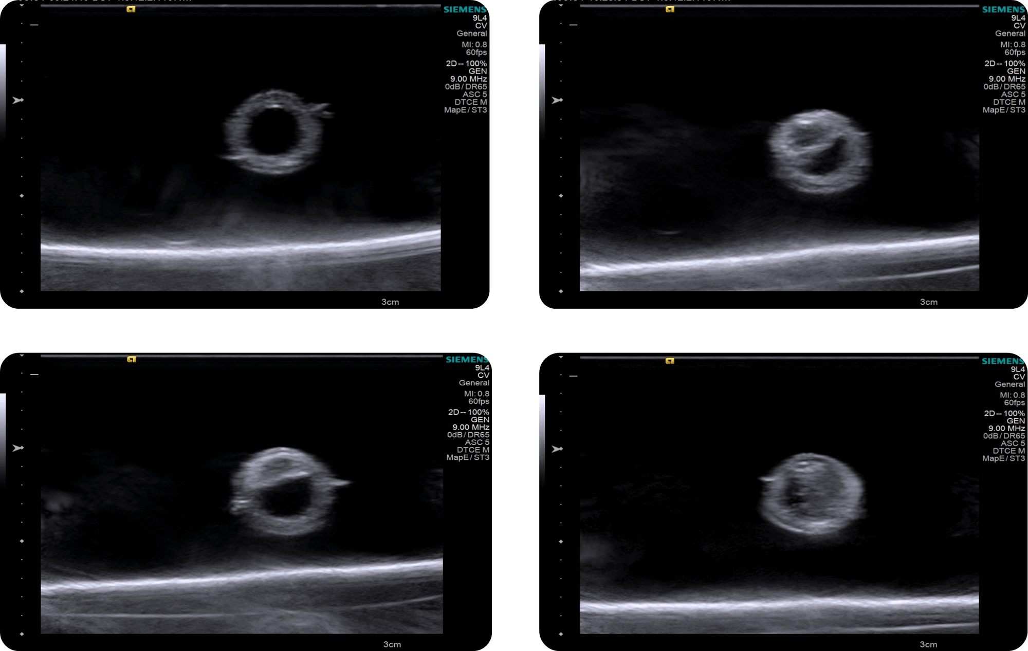
Figure 4: Inflation testing of arterial tissue phantoms with 0%, 25%, 50% and 75% stenosis.
This methodology was expanded to fresh human atherosclerotic plaques collected after surgeries.

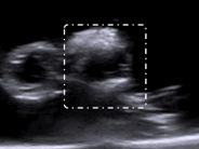
Figure 5: Inflation testing of fresh human atherosclerotic plaque combined with non-invasive ultrasound imaging. White rectangle on B-mode scan shows the branch inflated.
This framework combining non-invasive ultrasound imaging could allow for the identification of plaque features to decipher their proneness to rupture.